
Computed tomography
-
Vetriver Veterinary Clinic
Laski 24h, Latchorzew
-
State-of-the-art diagnostic imaging
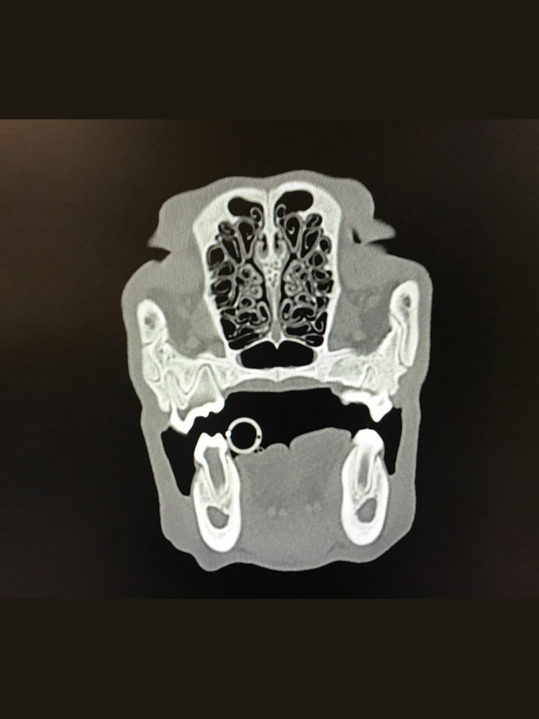
Examination by CT uses X-rays. With this examination, we obtain a number of cross-sections of the patient's body, and thus information that allows doctors to make a diagnosis, plan further diagnostic or therapeutic procedures. We achieve information that we are often unable to obtain during less advanced diagnostic imaging studies, such as X-ray or ultrasound.
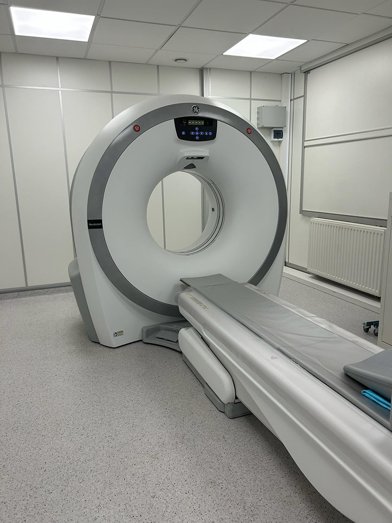
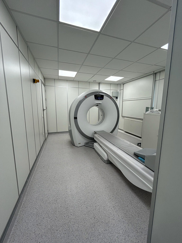
At our clinic we have a state-of-the-art 16-slice Revolution CT scanner, which provides even more precise imaging and reduces examination time, resulting in greater patient comfort and safety.
CT scanning is used to diagnose, among other things:
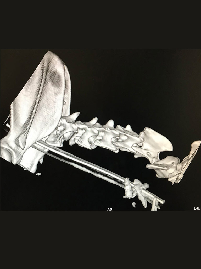
- diseases of the musculoskeletal system, including disorders of the spine,
- orthopedic disorders,
- diseases within the brain and head (including inflammations, tumors, post-traumatic conditions, congenital defects, diseases of small structures of the ear),
- diseases of the respiratory , digestive and urinary systems,
- to some extent diseases of the nervous system.
-
CT examination
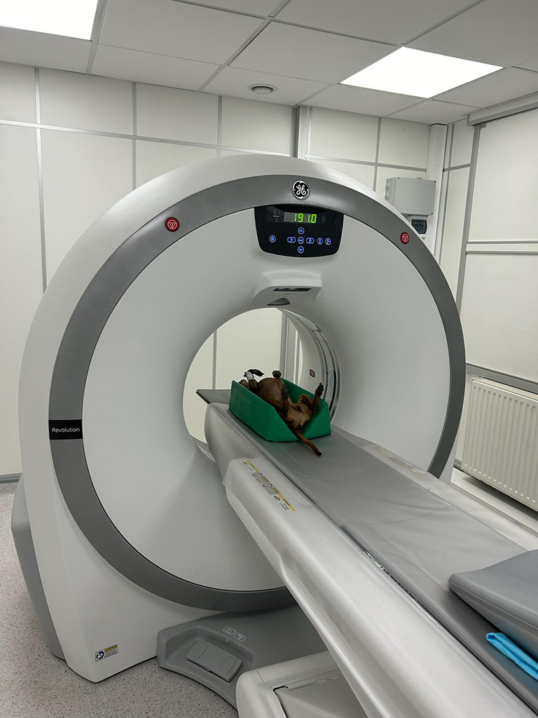
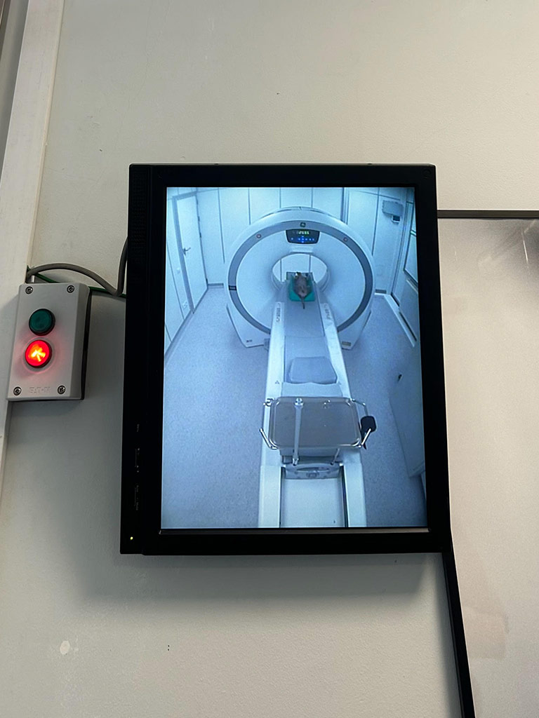
During the examination, the patient is placed on a moving platform, on which he or she rides inside the CT scanner. The animal must be under general anesthesia during the examination. During the examination, a lamp moves around the patient, which produces X-rays. The images produced during each rotation of the lamp around the patient's body are then summed up and reconstructed by a computer and, as a result, presented on a monitor as a picture of individual anatomical structures. A veterinary radiologist analyzes, interprets and describes the images obtained by the examination. The CT scan is a non-invasive and painless procedure.
 This site uses cookies for functional and statistical purposes